Bone Cross Section Under Microscope
Bone Cross Section Under Microscope. Be careful pushing it under the clips that the cover slide doesn't move or crack. Jump to navigation jump to search. From wikimedia commons, the free media repository. The jeol ion beam cross section polisher (cp) is widely used for preparing pristine samples prior to high resolution imaging and elemental analysis with the scanning electron microscope (sem). This slide showing a cross section of the mammalian trachea (wind pipe) contains examples of several different kinds of tissues. There are even muscles acting distal to forearm that attach on the humerus and cross multiple joints. The microscopic cross section represents the effective target area of a single target nucleus for an incident particle. Ladda ned bilder, illustrationer och vektorgrafik med cross section human med hög kvalitet till priser som passar projektets budget perfekt. The differences were significant in anterior.
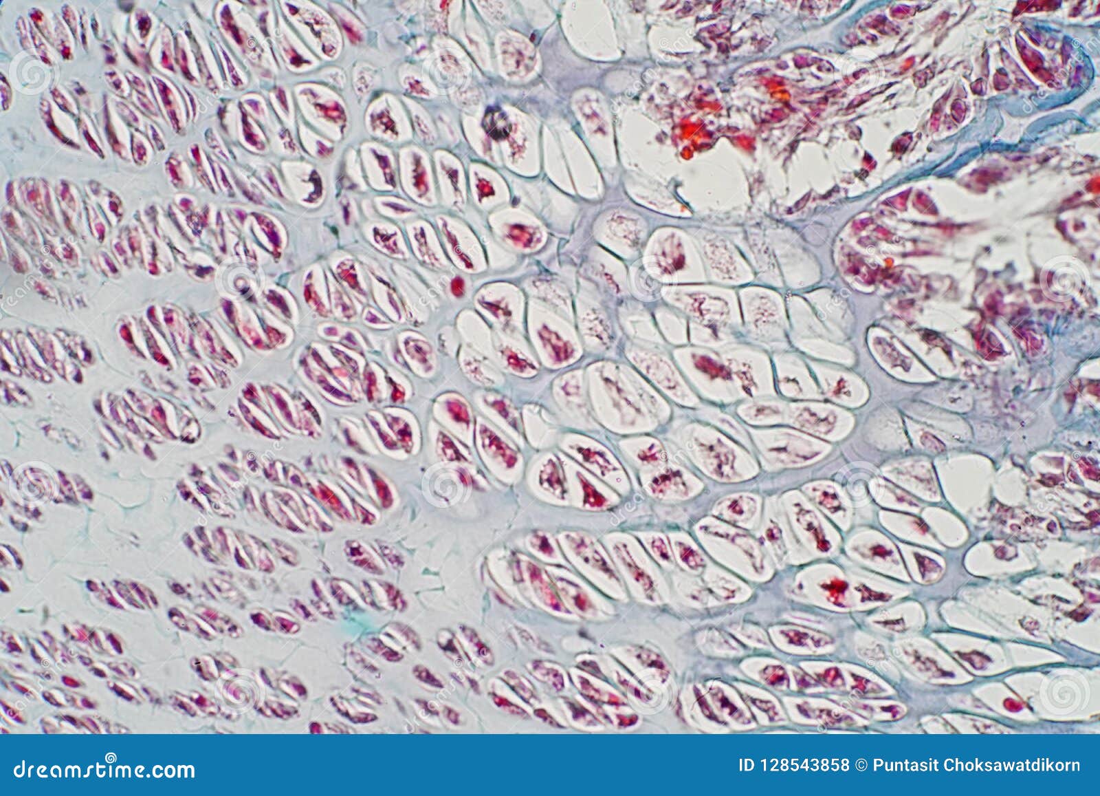
Monocot root cross section slide view under microscope for botany education. Huge collection, amazing choice, 100+ million high quality, affordable rf and rm images. Note that the bone matrix is deposited in concentric layers called lamellae. The major components of the cross section polisher (cp) are the ar ion source, shielding plate and specimen, as shown in fig. The large dark spots are passages for blood vessels and nerves. Trabecular bone found in metaphysis and epiphysis, as seen under microscope. The microscopic cross section represents the effective target area of a single target nucleus for an incident particle. From wikimedia commons, the free media repository.
The microscopic cross section represents the effective target area of a single target nucleus for an incident particle.
The nuclear cross section of a nucleus is used to describe the probability that a nuclear reaction will occur. Compact bone cross section courtesy: Like most sections of bone, it is strong, but it lacks the rigidity of the diaphysis. The jeol ion beam cross section polisher (cp) is widely used for preparing pristine samples prior to high resolution imaging and elemental analysis with the scanning electron microscope (sem). How to use a microscope. The large dark spots are passages for blood vessels and nerves. Where speed is essential, such as in surgical biopsies for cancer. Select the lowest power objective lens. When the light that enters the condenser is polarized by placing a polarizer in the filter holder and a second, crossed polarizer at the image plane. The edge of the shielding plate is positioned at the point where the cross section observation is desired, and the specimen is irradiated. The microscopic cross section measures the probability of occurrence of a particular nuclear reaction.
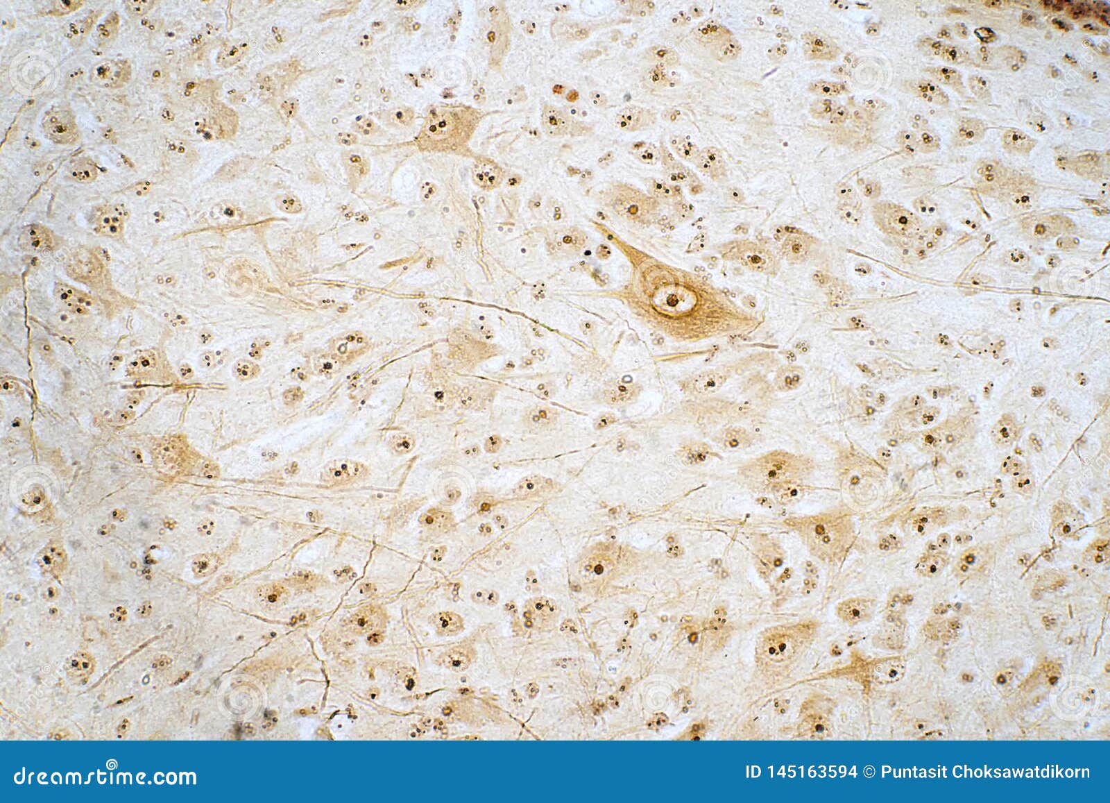
Jump to navigation jump to search. It is placed directly above a specimen. Bone marrow aspiration uses a hollow needle to remove a small sample (about 1 ml) of bone marrow for examination under a microscope. Note that the bone matrix is deposited in concentric layers called lamellae. Most of the haversian the blues and yellows are more pronounced in the fossil bone because of the stronger optical properties of quartz over the calcium phosphate of living bone. This simply involves placing a section of the bone on the microscope stage and viewing. The finished bone section will be bonded to a microscope slide and so the first step is to grind flat and polish the part of the bone that will be glued to the slide. This slide showing a cross section of the mammalian trachea (wind pipe) contains examples of several different kinds of tissues. These bone cells have long branching arms (d) which lets them communicate with. Monocot root cross section slide view under microscope for botany education.

They build the entire picture, improve your understanding, consolidate the information and facilitate recall.
They build the entire picture, improve your understanding, consolidate the information and facilitate recall. Find the perfect under microscope cross section cross stock photo. Huge collection, amazing choice, 100+ million high quality, affordable rf and rm images. Like most sections of bone, it is strong, but it lacks the rigidity of the diaphysis. There are even muscles acting distal to forearm that attach on the humerus and cross multiple joints. The finished bone section will be bonded to a microscope slide and so the first step is to grind flat and polish the part of the bone that will be glued to the slide. Be careful pushing it under the clips that the cover slide doesn't move or crack. The differences were significant in anterior. Bone cross section — stock image. Under the microscope footage of a transverse section of hard bone. Bone marrow is on the upper left. It is placed directly above a specimen. Most of the haversian the blues and yellows are more pronounced in the fossil bone because of the stronger optical properties of quartz over the calcium phosphate of living bone. The microscopic cross section represents the effective target area of a single target nucleus for an incident particle.
Bone marrow aspiration uses a hollow needle to remove a small sample (about 1 ml) of bone marrow for examination under a microscope. The differences were significant in anterior. Like most sections of bone, it is strong, but it lacks the rigidity of the diaphysis. The jeol ion beam cross section polisher (cp) is widely used for preparing pristine samples prior to high resolution imaging and elemental analysis with the scanning electron microscope (sem). Compact bone cross section courtesy: The lining of the trachea consists of a type of this slide contains a section of dried compact bone.
The major components of the cross section polisher (cp) are the ar ion source, shielding plate and specimen, as shown in fig.
The jeol ion beam cross section polisher (cp) is widely used for preparing pristine samples prior to high resolution imaging and elemental analysis with the scanning electron microscope (sem). Where speed is essential, such as in surgical biopsies for cancer. The finished bone section will be bonded to a microscope slide and so the first step is to grind flat and polish the part of the bone that will be glued to the slide. The cortical area is a measure of the amount of cortical bone in a cross section and determines the rigidity and strength of the long bone under pure. Thin section of dinosaur bone. The major components of the cross section polisher (cp) are the ar ion source, shielding plate and specimen, as shown in fig. Jump to navigation jump to search. Thin section of dinosaur bone. If you were to look at it in under a microscope, it would. Huge collection, amazing choice, 100+ million high quality, affordable rf and rm images.
The edge of the shielding plate is positioned at the point where the cross section observation is desired, and the specimen is irradiated bone cross section. This simply involves placing a section of the bone on the microscope stage and viewing.

Posting Komentar untuk "Bone Cross Section Under Microscope"